
Resources
Equipment
-
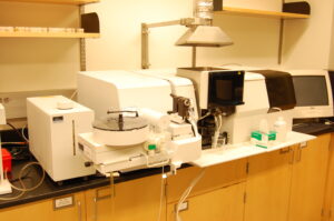
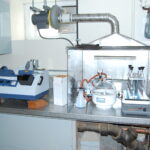
Location: Morley Science Room 236 About 70 different chemical elements can be quantitatively determined using the Atomic Absorption Spectrometer (AAS). The AAS very accurately measures the absorption of a specific wavelength of light by atoms that have been converted to the gaseous state with either a 2300º C acetylene flame, or utilizing a temperature step program and a graphite tube. Most recently the AAS has been used to measure the amount of common metals in water, lead in river sediments and local house paint, and iron in alpine soils.
-
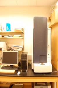
Location: Morley Science Room 230 The MALDI allows for very high resolution mass analysis of samples. The MALDI can analyze very large molecules up to several hundred thousand Daltons. In the instrument, molecules are ionized using a combination of a high powered laser and a matrix molecule. The charged particles are accelerated toward a detector in an electrostatic field. The mass is measured by the length of time needed for a molecule to arrive at the detector approximately 1 meter away. This instrument is most commonly used to identify proteins based on their characteristic digestion “fingerprint”.
-
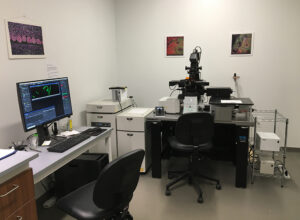
Location: Hopper Science Room G13 The Nikon High Definition Resonant Scanning Confocal A1rHD looks at fluorescently stained materials and can localize cell components or other areas of interest on biological samples. For example, the confocal can look at live anesthetized tiny fish embryos and make a movie of the heart beating and the valves opening and closing. It can also help assess the properties of various materials.
-
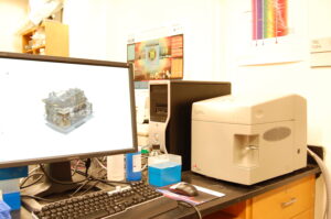
Location: Thompson Biology Room 108 The flow cytometer is used to analyze physical and biochemical properties of living cells. The instrument draws cells into a flow cell where they scatter the light from a laser. The degree of scatter gives information about the size and internal complexity of the cells. In addition by labeling internal molecules (typically proteins) in the cell with fluorescent chemicals, we can obtain information about cell function. Our instrument uses 3 different lasers allowing us to simultaneously examine several proteins at once.
-
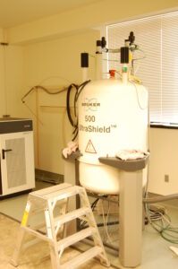
Location: Morley Science Room 154 Model: Bruker Ultrashield 500MHz
This instrument analyzes the structure of molecules by evaluating the electromagnetic radiation emitted by certain atomic nuclei when they are subjected to their electromagnetic resonance frequency in a strong magnetic field. Our Brüker Avance DRX 500 MHz NMR spectrometer is equipped with a Z-axis gradient unit and a variable temperature controller, and can be operated with either a 5mm broad-band multinuclear (PABBO) probe or a 5mm 1H/13C/15N (TXI) inverse probe. This instrument allows chemical scientists at Williams to determine the structures of molecules from a wide variety of projects, ranging from natural product total synthesis, polymer synthesis, materials science and catalyst development research projects, natural product isolation studies, and structure determination of small peptides.
Contact: Silas Brown [email protected]
-
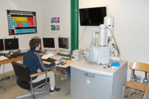
The SEM uses a focused electron beam to obtain very high resolution 3 dimensional images of the surface topography of virtually any sample that will fit into the scope. In addition, we can use the SEM to obtain information about the elemental composition of a sample.The electron microscopes and other scopes are located in the Oberndorf Family Microscopy Suite on the ground floor of Hopper Science Center – Rooms G 08 thru G13.
-
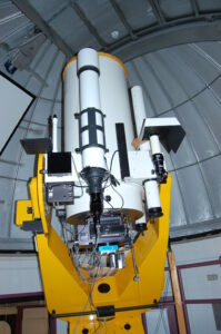
The observatory houses a professional-grade 24-inch (0.6-m) Cassegrain reflecting telescope from DFM Engineering. It is principally used in introductory and advanced courses for imaging using a FLI PL 16803 CCD camera and a variety of optical filters. It also carries an Optomechanics C-10 spectrograph, recently upgraded with new components and a new Apogee U1107 spectroscopy CCD. The telescope is also used to conduct research on variable massive stars in young star clusters, and in addition, to observe occultations of stars by small bodies in the outer solar system.
-
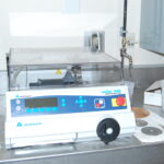
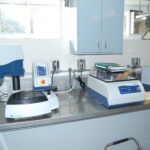
Equipped with high-precision, diamond-based cutting, grinding and polishing equipment, the thin sectioning lab prepares solid materials for analysis. Typical preparation creates “thin sections”, samples mounted to glass microscope slides, then ground to a thickness of 30 microns or less. At this thickness, most solid materials transmit light and can be analyzed with an optical microscope. Thin sections are also suitable for scanning electron microscope (SEM) analysis. The lab also produces “bulk mounts”, whole samples set in resin with one side cut and polished. Bulk-mounts are typically analyzed by SEM.
-
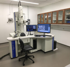
Hopper Science Center room G08 The TEM uses a focused electron beam that can examine sectioned samples at high magnification and resolution. The fine structure of biological materials as well as nanoparticles and viruses can all be viewed at magnifications up to 500,000x.
Science Shop
The Hopper Science Shop is a resource for all faculty, staff, and students. We support faculty research projects, classroom demonstrations and independent studies by designing, fabricating, modifying or repairing individual parts or complete systems. The capabilities of the shop include machining and fabrication with metals and plastics, laser cutting and engraving, CNC milling, 3D printing, woodworking, welding and electronics fabrication. Students may use Science Shop facilities for approved independent study, and on a space-available basis for individual projects.
There are a limited number of work-study opportunities for students.
For more information contact:
- Jason Mativi – Senior Science Center Shop Engineer (jwm1@williams.edu, x2205)
- Dave Williams – Science Center Shop Engineer (djw10@williams.edu, x2230)
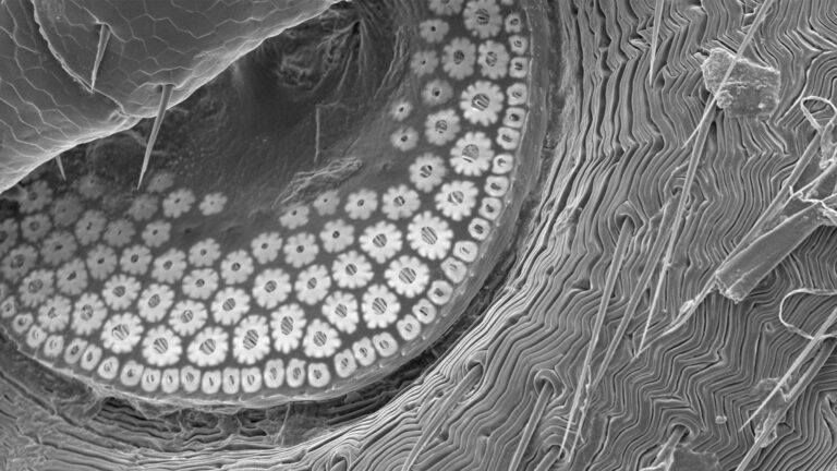
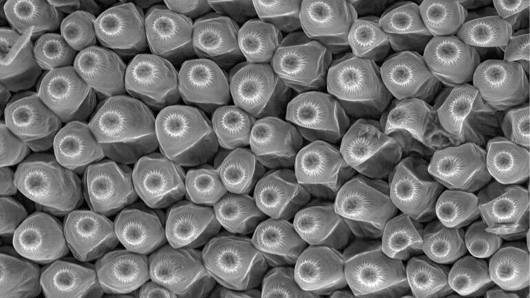
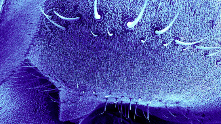
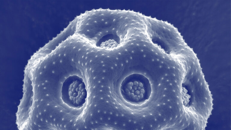
Electron Microscopy Lab
The Electron microscopy Lab is a multidiscipline resource for students and faculty located in the Hopper Science building. Students can do thesis research or semester research using both the scanning electron microscope (SEM) and the transmission electron microscope (TEM). Thesis work has included geology students looking at the morphology and chemistry of thin sections of rock on the SEM. Labs for courses in geology, chemistry and biology use it to examine specimen samples. For example, biology students have done a study of fish spinal cords and the fine structure of nerves in the TEM, and chemistry labs used the SEM to look at nanoparticles in the sub-micron range. Summer Science students use the SEM to look at an assortment of samples from blood cells to hair to microchips. Winter Study classes also utilize the SEM and TEM. Students are trained to do their own sectioning for the TEM, and obtain both microscope slides for the light microscope as well as thin sections for the TEM for higher magnifications. A technician is on hand to assist students and faculty for both specimen prep and using the microscopes.
Electron microscopy lab photos
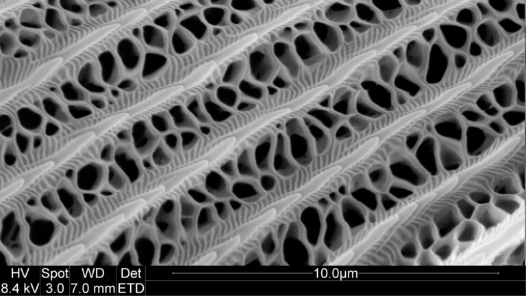
Tiger Swallowtail Wing
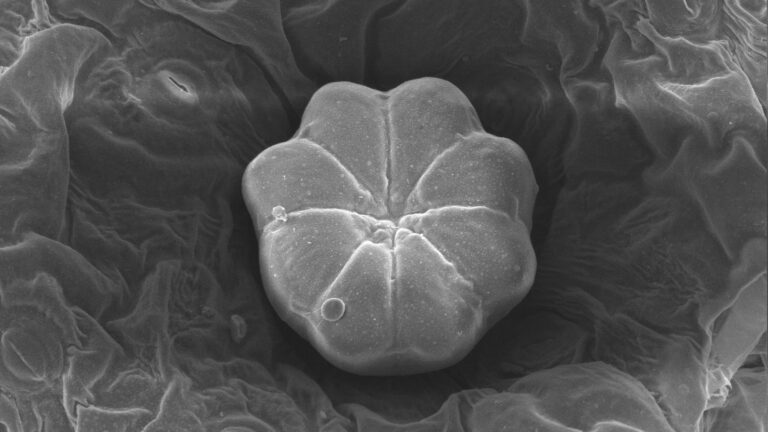
Underside of a Tick
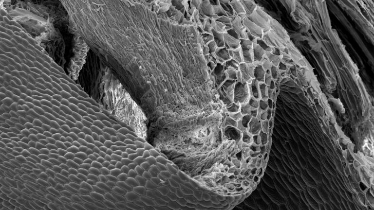
Swedish Ivy Leaf
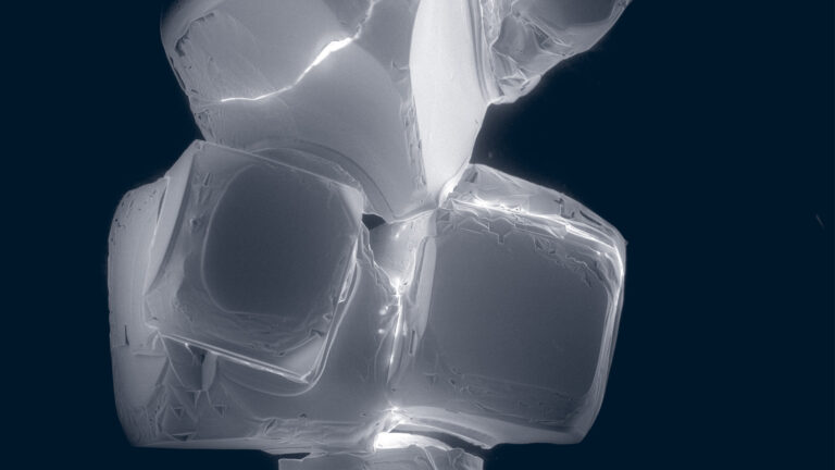
Recrystallized Salt
Orchid
Geranium
Fly Spikes
Chickweed Pollen
Faculty Funding Resources
Internal Funding Links:
Divisional Research Funding Committee (DRFC) Request Forms:
Now available as Google Forms (must be logged into Google with your williams.edu email address).
- Research Assistant Application AY 2025-26 (please complete whether requesting DRFC funding or if you have a grant)
- Discretionary Funds Application FY26
- Larger Research-Related Application FY26
- Summer Science Research 2025 Application
External Grant Links:
Science Center
31 Morley Drive
Williamstown, MA 01267
Phone: 413-597-2167